Your email address will not be published.


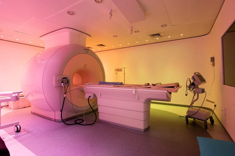
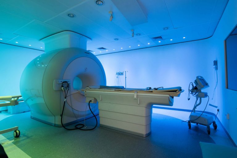
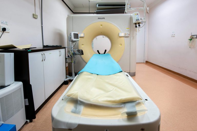
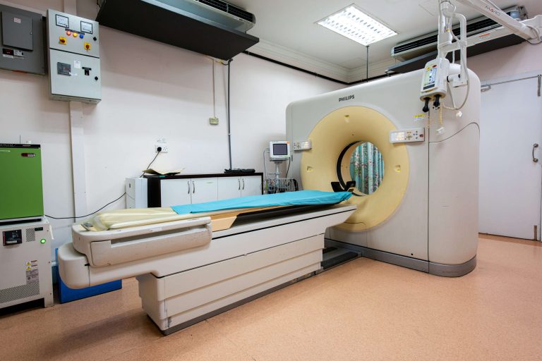
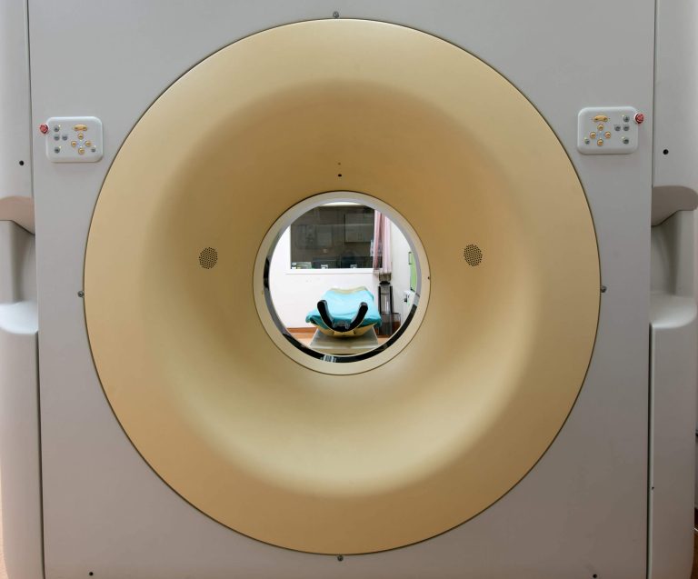
Putra Specialist Hospital Melaka offers a full-service of CT Scan from routine to Interventional radiology to look at the bone or tissue disorder.
We also offer calcium scoring test, a procedure to determine if the coronary arteries are blocked or narrowed by the build-up of plaque – an indicator of atherosclerosis or coronary artery disease (CAD). The information obtained can help evaluate if you are at increased risk for heart attack.
These highly-specialized imaging tool has a 64-slice scanner to create cross-sectional images of bones, soft tissues, blood vessels and organs. The scan allows doctors to see the inner structure and more detailed images of the body.
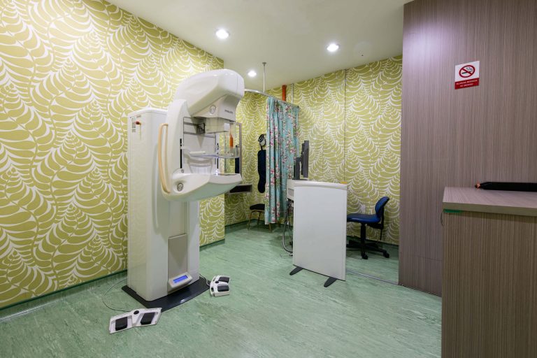
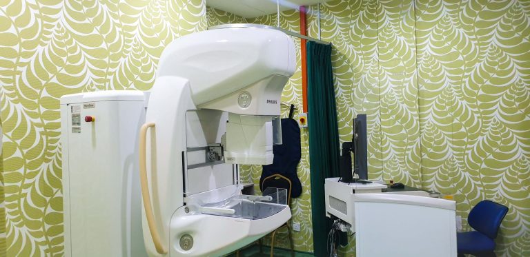
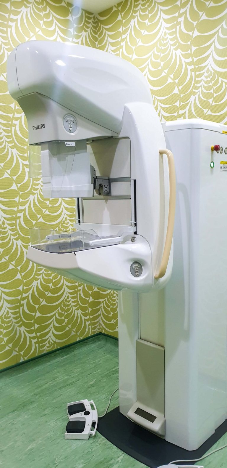
Mammography remains the gold standard for screening of early stage breast cancer. Putra Specialist Hospital Melaka is the only centre in Melaka which offers Micro-Dose Digital Mammography.
Micro-Dose Mammography provides less stressful mammography experience because it can give warm, curved patient support. It is also easy positioning and therefore allows faster imaging time.
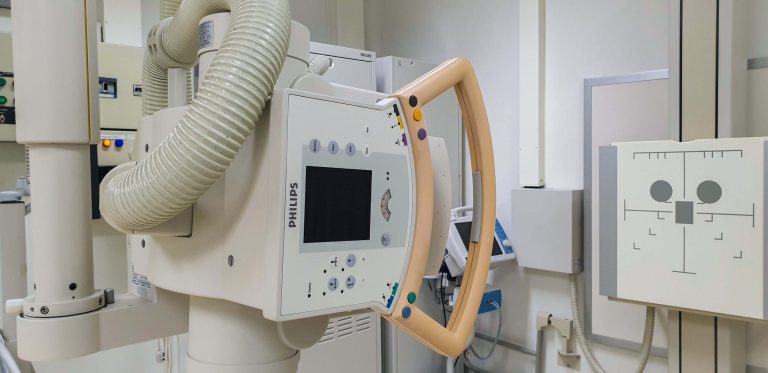
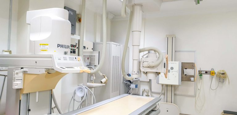
Fluoroscopy, like most other imaging tests, can be used to see what’s going on within our bodies. The images that the procedure provides can be helpful in diagnosing abnormalities or in treating these abnormalities.
Fluoroscopy exams can provide detailed “moving’ images of entire body systems, including the skeletal, digestive, urinary, respiratory, and reproductive systems, or it can look at specific body organs, such as the heart, lungs, or kidneys.
The moving images allow to see if there is a blood clot in the veins or arteries, if the bones are healthy, or if the digestive tract is working properly. One common fluoroscopy exam involves a barium (or contrast) swallow, which passes through the GI tract and allows one to see the GI movements in even greater detail.
Fluoroscopy can be used in any of the following situations:
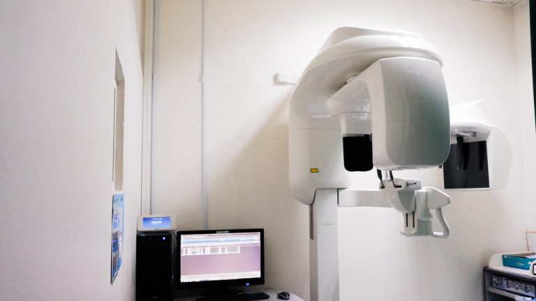
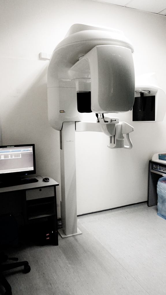
Orthopantomogram also known as OPG is a panoramic single image radiograph of the mandible, maxilla and teeth. It is often encountered in dental practice and occasionally in the emergency department; providing a convenient, inexpensive and rapid way to evaluate the gross anatomy of the jaws and related pathology.
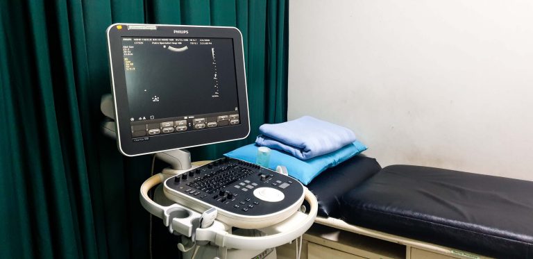
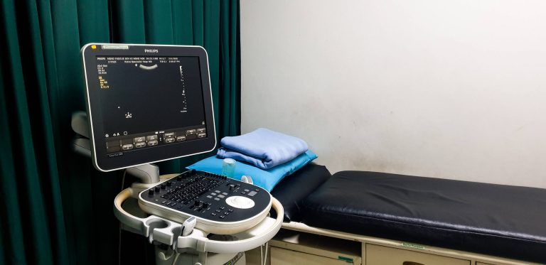
Ultrasound uses sound waves to produce pictures inside of the body. It is used to help diagnose the causes of pain, swelling and infection in the body's internal organs and to examine a baby in pregnant women and the brain and hips in infants. It's also used to help guide biopsies, diagnose heart conditions, and assess damage after a heart attack. Ultrasound is safe, non-invasive, and does not use ionizing radiation.
An X-ray is a quick, painless test that produces images of the structures inside your body - particularly your bones. X-ray beams pass through the body, and they are absorbed in different amounts depending on the density of the material they pass through. Dense materials, such as bone and metal, show up as white on X-rays. The air in lungs shows up as black. Fat and muscle appear as shades of grey.
The most familiar use of x-rays is checking for fractures (broken bones), but x-rays are also used in other ways. For example, chest x-rays can spot pneumonia.
Related Photos
Your email address will not be published.
© 2024 Putra Hospital Melaka. All Rights Powered by IT Department KKLIU: 2346 / EXP 31.12.26